-2700x842-1920w.png)


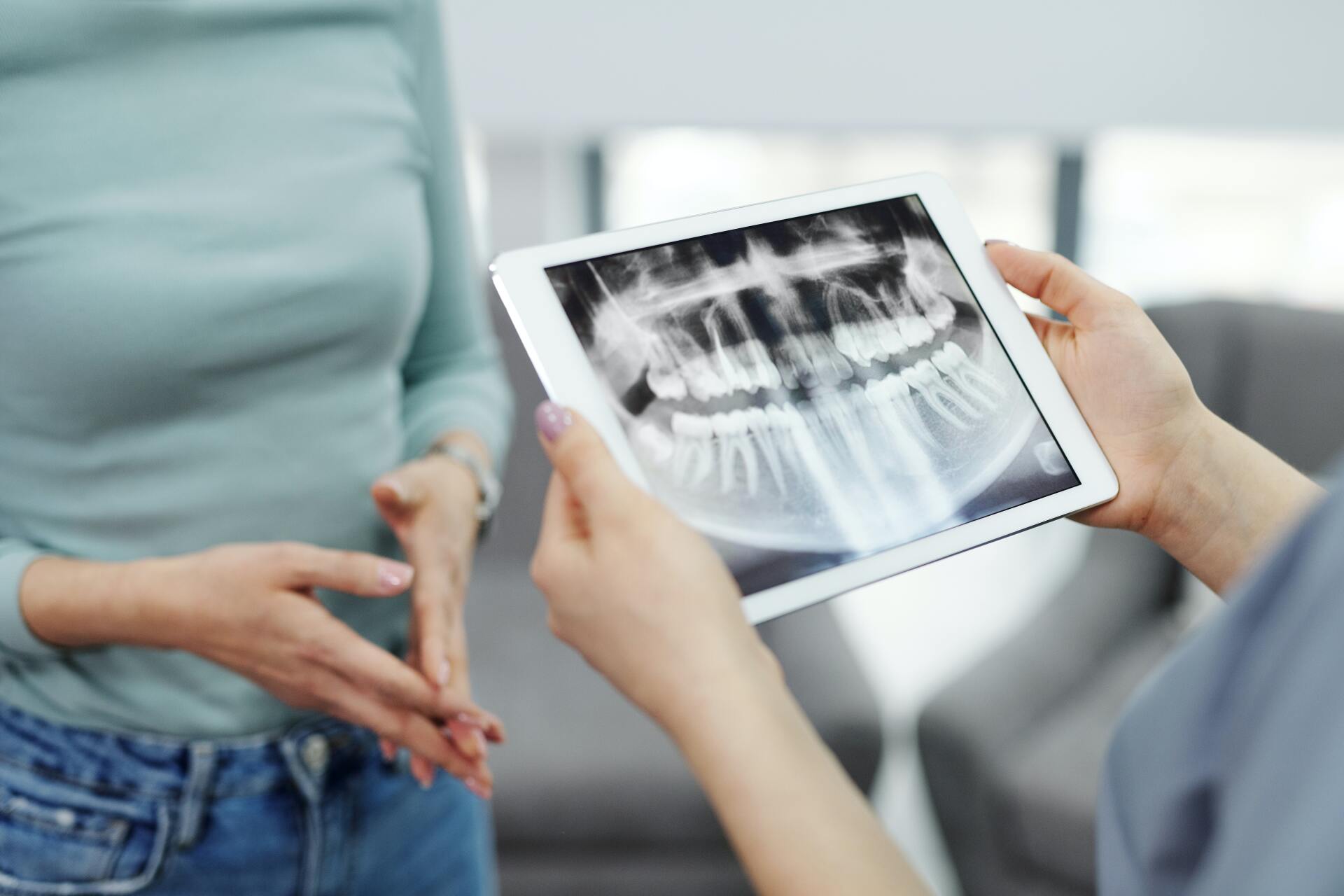
Imaging Options for Diagnosing TMJ
The temporomandibular joint, or TMJ, is an anatomically complex joint, composed of muscles, joints, ligaments, bones, and discs. Proper jaw movement requires a high level of interaction and coordination among all these components, especially the articular disc (which absorbs stress), the adductors (jaw-closing muscles), the abductors (jaw-opening muscles), and jaw ligaments. This makes diagnosing problems or dysfunction with the TMJ extremely difficult.
Properly diagnosing disorders of the TMJ—collectively referred to as TMD—requires both clinical examinations and medical imaging in order to develop an accurate assessment of the joint, its function, and its surrounding bone and tissue.
If you are experiencing pain, discomfort, or dysfunction in your TMJ, you will first have a clinical evaluation. This is when your doctor or TMJ specialist will discuss your symptoms with you and examine your jaw, pressing on areas around it to identify any sensitivity. He or she will also observe the range of motion of your jaw and listen to and feel around your jaw while you open and close your mouth.
In some cases, the doctor’s clinical findings are enough to diagnose a TMD and begin treatment. Other times, additional diagnostic imaging will be necessary to confirm the presence of a TMD.
What is The Role of Imaging in Diagnosing TMJ?
Diagnostic imaging allows a TMJ practitioner to better assess the various bones, tissues, and muscles of the TMJ, including their health and how the hard and soft tissues are working together in the joint. Imaging also enables the doctor to evaluate the effectiveness of current treatment, as well as to confirm the extent or progression of joint disease.
Your doctor, dentist, or TMJ specialist may recommend additional imaging if:
- Your treatment plan to date has failed, or if your symptoms are getting worse
- You have a history of trauma or TMD
- Significant dysfunction, infection, or abnormality is suspected
- You’ve had significant changes in occlusion (the alignment of your teeth and jaws)
- You are experiencing sensory or motor abnormalities
- Your TMD treatment plan includes orthodontia or extensive reconstructive work
However, because the TMJ is so complex, it can be hard to capture all the aspects of it with certain imaging, or even with only one type of imaging.
What Types of Imaging Are Used for Diagnosing TMJ
The type of imaging a TMJ doctor will use depends on:
- Whether it’s hard or soft tissue (or both) that will be imaged
- Cost
- Availability of imaging technology or method
- Radiation dose requirements or limitations
- The amount of diagnostic information required (different methods provide different levels and types of imaging data)
In general, there are three types of imaging used to diagnose TMJ. While some methods have advantages over others, TMJ specialists will often use a combination of all three to create as comprehensive a picture as possible of your condition. Not only does this allow the provider to better pinpoint the cause of your symptoms and any other factors at play, but it also enables him or her to develop a better treatment plan for optimal results.
The three types of imaging are:
- Radiography, such as dental x-rays, for examining teeth and the jaws
- Computed tomography, or CT scans, for detailed images of the bones within the TMJ
- Magnetic resonance imaging, or MRI, for identifying problems with the joint’s disc or surrounding soft tissue.
Radiography and CT scans are typically used first to evaluate the hard tissue and build a conservative treatment plan. If symptoms don't respond to treatment, or if the doctor's clinical findings suggest disc displacement or significant TMJ pain or dysfunction, then the soft tissues will be examined using an MRI or arthrography.
What Are the Best Imaging Methods for Diagnosing TMJ?
Every diagnostic method has advantages and disadvantages and can capture a different aspect of the TMJ. Because of the joint's complexity, and the limitation of any single imaging method, your doctor or TMJ specialist may order a combination of imaging tests for a more accurate assessment.
For hard tissue evaluation, your doctor may use:
Conventional radiography like X-rays, panoramic x-rays, or conventional tomography. While not the preferred method, conventional radiography—particularly tomography—is good as a general screening tool for an initial assessment. It's best for broad visualization and can identify asymmetry, tumors, trauma, or degenerative bone changes, but only in late stages.
Conventional radiography has significant limitations, however. A thorough understanding of the TMJ and its functional status requires a 3D view. Radiography only produces 2D images that are on a flat plan and slightly distorted as a result of the bony structures that are over the TMJ. While conventional tomography is better—it replaces traditional plain film techniques and can show the TMJ in thin slices or layers—films have to be taken in different places to accomplish the necessary detail for proper diagnosis and treatment planning, making it time-consuming and more susceptible to processing errors.
- Pros: Radiography is widely accessible, produces fast imaging, is cost-effective, and delivers lower doses of radiation than other methods.
- Cons: Radiography only produces 2D images of the TMJ and can only show the bony elements of the joint. Conventional tomography can be more time-intensive than other methods or even computed tomography.
Best for: Initial assessments of TMJ disorders.
Computed tomography (CT). Computed tomography is essentially digital tomography, which uses a computer to assemble a series of 2D digital x-rays into a 3D image that shows the relationship among different components in the TMJ, including both the bony elements and adjacent soft tissues.
CT scans are better than conventional radiography and produce a clearer picture, but they still only image the TMJ in 2D, making them more suitable for use in orthodontics and for neutral angles of the head, teeth, jaw, and neck.
- Pros: CT is good for 3D reconstruction and assessing bony structures. Improved contrast resolution makes tissue with small differences in density more distinguishable, and data from one imaging study can be viewed in various planes as well as in 3D. There is also no superimposition of bony structures outside the area of interest, as there is in traditional radiography.
- Cons: CT often comes at a higher cost and with higher radiation dosages than other methods. Images are susceptible to distortions from metallic objects like restorations or prostheses, and the TMJ disc and other soft tissues are usually not visible.
Best for: Detailed assessment of bony structures and disorders.
Cone beam CT. Cone beam CT, or CBCT, is an advancement of computed tomography that is well suited for imaging the hard tissues of the skull and jaws. It provides a high-resolution multiplanar reconstruction of the teeth, mandible, airway, sinuses, skill, TMJ, and cervical spine in 3D sections, all with low doses of radiation.
Cone beam CT is better than conventional CT because large areas can be imaged in a single scan and then reformatted into multiple planes, allowing for more detailed views of the TMJ, skull base, and the rest of the jaw.
- Pros: CBCT produces lower radiation than a medical CT and produces a lot of data in minutes. Like conventional CT, CBCT doesn't experience the superimposition of bony structures, but, unlike CT, there is no distortion from metallic objects.
- Cons: CBCT has limited views of soft tissue and disc placement, and it isn’t a preferred method for patients who can’t remain motionless during the scan.
Best for: 3D images of teeth, nerve pathways, bone, and soft tissues in a single scan, especially when regular x-rays are insufficient.
If your doctor or TMJ specialist determines you need additional imaging of soft tissues, you may receive one of the following types of diagnostic imaging:
Magnetic resonance imaging, or MRI. MRIs are considered "the gold standard" in imaging the soft tissue component of the TMJ. Instead of radiation, MRIs use magnetic fields and radiofrequency pulses to produce multiple digital image slices, allowing the doctor to examine the articular disc in terms of location and possible displacement. This information can help detect early signs of TMD and joint effusion (a collection of fluid in the TMJ space), as well as signs of inflammation or swelling in the joint.
- Pros: MRIs can produce high-resolution and tissue-contrast images of the jaw in both the open and closed position with no radiation.
- Cons: MRIs are one of the most expensive methods of imaging technology and take more time to conduct. They also provide a limited ability to assess bony changes.
Best for: Assessing soft tissue and disc placement, as well as evaluating the anatomy and jaw biomechanics of the jaw and TMJ in both the open and closed positions.
Ultrasonography/diagnostic ultrasound. Ultrasonography is a recent technology that can help your doctor get a sense of the TMJ's motion and disc position. It's an inexpensive and noninvasive way to conduct a "real-time evaluation" of disc positioning in the open-mouth position. However, it's still new, and the value of the data and information produced is highly dependent on the examiner's skill and the type and quality of equipment used.
- Pros: Ultrasonography can be performed quickly and safely in a practitioner’s clinic. It’s inexpensive and noninvasive.
- Cons: The quality of imagery and assessment of ultrasonography results relies heavily on the skill of the examiner.
Best for: Initial screening for TMD and as a supplement to clinical evaluation.
A less-often, least-preferred method of diagnosing TMJ is arthrography. Arthrography is an invasive imaging technique that uses a radiopaque iodine-based agent injected into one or both TMJ spaces to take a radiograph. Although it can detect small perforations of the disc or attachments, arthrography can’t detect medial disc displacement and the procedure can result in post-operative discomfort as well as an allergic reaction to the contrast agent.
Treat Your TMJ with Diagnostic Imaging from REstore TMJ & Sleep Therapy
If you or a loved one have been suffering from TMD or pain in your TMJ, relief can’t come soon enough. Diagnostic imaging for TMJ can help provide a comprehensive assessment of your condition so you can receive the best, most effective treatment possible.
Dr. Katherine S. Phillips of Restore TMJ & Sleep Therapy is a double-board certified TMJ specialist who has spent more than a decade diagnosing and treating TMJ and sleep disorders, helping thousands of patients with TMD live a healthier life free from pain.
Call Restore TMJ & Sleep Therapy today at 281-296-6797 to schedule your no-obligation consultation. With diagnostic imaging, we can treat the symptoms and the cause of your TMJ pain for results that last.
Contact Us Today For A Consultation!
Contact Us


REstore TMJ & Sleep Therapy P.A.
1001 Medical Plaza Drive,
Suite 200 | The Woodlands, TX 77380
281-296-6797


Dr. Phillips serves TMJ & Sleep patients in: The Woodlands TX | Spring TX | Conroe TX Tomball TX | Cypress, TX | Houston, TX | Kingwood TX | Humble, TX | Katy TX
© 2023 by REstore TMJ & Sleep Therapy | Terms Of Service & Privacy Policy | XML Sitemap